Electronics, Opto-electronics, Sensing and Biomedical Devices
Activities within this Focus Area include: developing energy-efficient, high-performance electronic devices and systems using two-dimensional layered materials and heterostructures of nanocarbons and transition metal chalcogenides for computation, as some examples; electronic and opto-electronic devices for enhanced detection of radiation from the IR, visible, to the UV regimes; developing novel techniques using laser holography for the nanofabrication of 3D photonic crystals; physical and chemical sensors to detect biothreats and toxins for defense and homeland security, including in extreme environments; and biomedical devices for healthcare, that may also be implantable and biocompatible.
Electronics, Opto-electronics and Photonics
As example of activities in this area, Research in PACCAR Director’s lab, the Nanoscale Materials and Devices Lab, Prof. Kaul’s group has designed, fabricated, and characterized multiple photodetector devices, please refer to NMDL site for the latest publications in this area from the Kaul Group.
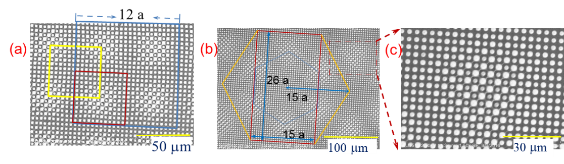
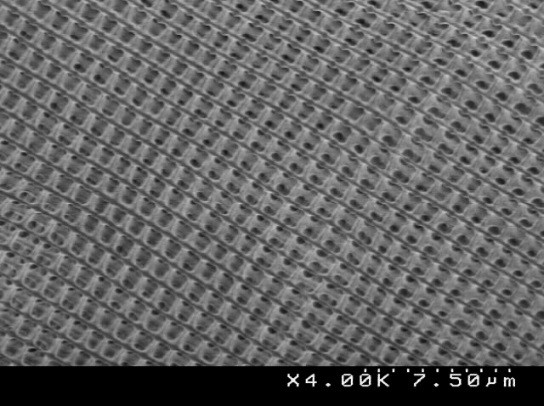 Similarly, PACCAR-affiliated faculty Prof. Yuankun Lin’s research lab, the Photonic Band-gap Materials Lab, have efforts in the fabrication of 3D photonic crystals formed by a simplified method of laser holographic fabrication. The SEM of the newly discovered graded photonic super-crystals is shown on the right with a square lattice in a square cluster. Shown below in a) is the square lattice in a hexagonal cluster, and in (b) a large-area image of the structure is depicted, while in (c) a magnified region is captured from the area within the dashed red rectangular region in (b).
The developed technique grants flexibility in designing 2D photonic lattices with size-graded features, differing periodicities, and differing symmetries, as well as lattices having simultaneously two periodicities and two symmetries in high resolution. Photonic cavities can also be generated in the graded super-lattice simultaneously through a laser one-exposure process. A high quality factor of over 1.56×10⁶ for a cavity mode in the graded photonic lattice with a large super-cell is predicted by simulations for low threshold lasing application.
References:
D. Lowell, J. Lutkenhaus, D. George, U. Philipose, B. Chen, and Y. Lin, “ Simultaneous direct holographic fabrication of photonic cavity and graded photonic lattice with dual periodicity, dual basis, and dual symmetry,” Opt. Express 25, 14444-14452 (2017).
D. Lowell, S. Hassan, M. Adewole, U. Philipose, B. Chen, and Y. Lin, “Holographic fabrication of graded photonic super-crystals using an integrated spatial light modulator and reflective optical element laser projection system,” Appl. Opt. 56, 9888 (2017).
D.Lowell, S. Hassan, O. Sale, M. Adewole, N. Hurley, U. Philipose, B. Chen, and Y. Lin, “Holographic fabrication of graded photonic super-quasi-crystals with multiple-level gradients,” Appl. Optics 57, 6598 (2018).
Sensing and Biomedical Devices
The need for low-cost healthcare and disease diagnostics is a grand challenge area which includes: the development of implantable, biocompatible devices for sensing and detection of pathogens; biophotonic devices; cancer imaging and therapeutics using nanomaterials; neural probes and electrodes for the brain initiative; sensors for various biomarkers such as pH, antigen-antibody interactions, protein-antigen interactions; and microfluidic devices for lab-on-chip. We describe some of our activities aligned with the directions of PTI in this focus area.
Retinal Prosthesis
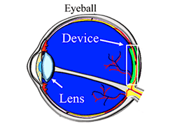
PTI-Director Kaul’s research lab, The Nanomaterials and Devices Lab (NMDL), is exploring devices for retinal degenerative diseases such as age-related macular degeneration (AMD). In AMD a continuous degeneration of photoreceptors in the retina is experienced, which can eventually lead to complete blindness. The photoreceptor layer of the retina malfunctions while the inner nuclear layer and the ganglion cells remain fully functional. Pharmaceutical treatments have been developed in an attempt to slow down tissue degeneration, but no method to halt or completely retard the progression has been successfully developed. Retinal prosthetic devices may be viable as a vision-recovery methodology to restore vision through electrical stimulation. Another approach to combat AMD is via the use of artificially implantable photodetectors that are physically placed on the retina. This approach, while promising, suffers from issues of Si substrate rigidity, where retinal cells migrate to fill the empty space between the implant and retinal pigment epithelium (RPE), which leads to the formation of scar tissue and cell reorganization in the various layers of the retina. Having large format photodetector pixels that are flexible and conformable allows the implant to be in intimate contact to RPE, and alleviates the issues faced with rigid and planar Si implants. Besides this advantage, such conformable structures also have the potential to increase areal coverage where photodetector pixels can be constructed to enhance the field of view by collecting more of the incoming radiation and focusing it onto the retina, given the naturally curved, hemispherical geometry of the eye.
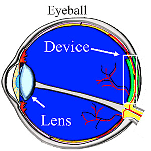 In Prof. Kaul’s research group, a new approach was utilized to form photosensitive pixels that utilize heterostructures of ink-jet printed two dimensional (2D) layered materials, specifically photosensitive and semiconducting molybdenum disulfide (MoS2) and electrically conducting graphene, using inks that also show a high degree of biocompatibility as gauged via cell proliferation tests. In addition, the biocompatible inks are formed on flexible and conformable substrates using low-cost, additive manufacturing technique, specifically ink-jet printing, which increases the likelihood of high-throughput translational opportunities targeted specifically toward AMD. Shown on the right is a representation of where such devices maybe implanted within the curved surface of the eye.
Reference:
R. Hossain, I. Deaguero, T. Boland, and A. B. Kaul, “Large-format, biocompatible, ink-jet printed 2D-heterostructure photodetector on flexible substrates,” Nature npj 2D Materials and Applications Journal, 1(28) (2017); DOI: 10.1038/s41699-0170034-2
Wireless Power Transfer (WPT) System Design for Optogenetic Implants
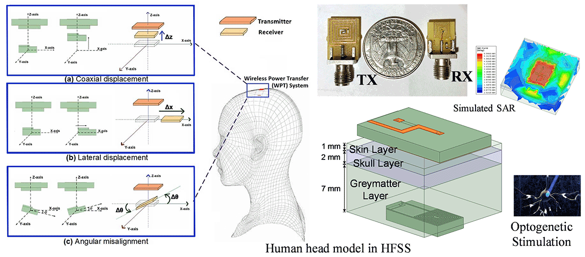
PACCAR-affiliated faculty, Prof. Ifana Mahbub and her research group, The Integrated Biomedical Circuits and Systems Laboratory (iBioCASL), is working on optogenetics which is a clinical methodology that stimulates genetically modified neurons in the brain using Lasers or µLEDs. The most challenging part of designing an optoelectronic implant is to efficiently deliver power to the implant despite the loose coupling between the transmitter (TX) and receiver (RX) coils because of the various displacements (coaxial, lateral and angular etc.). Dr. Mahbub and her group is analyzing and modeling the effects of various displacements on the link-efficiency and propagation loss though the brain tissue model which consists of the skin, skull and grey matter layer and verified the theoretical modeling with fabricated spiral coils on FR4 substrate and bovine muscle as the tissue media, as depicted in the figure below. Phantom human heads are being developed so that the dielectric parameters are similar to the real-life scenario. The modeling and empirical validation also helps to develop a closed-loop power regulation system to instantaneously compensate for the additional losses due to loose-coupling.
Reference:
D. K. Biswas, N. T. Tasneem, I. Mahbub, "Modeling the effects of displacements on link efficiency and propagation loss for a miniaturized wirelessly powered optogenetic implant" in IEEE Journal of Electromagnetics, RF and Microwaves in Medicine and Biology. (under review)
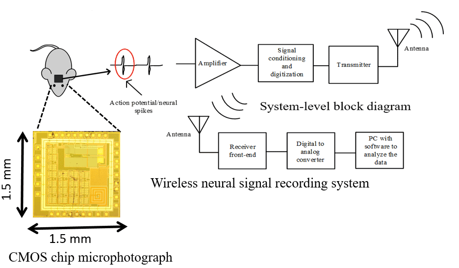
Wireless Neural Signal Recording System
PACCAR-affiliated faculty, Prof. Ifana Mahbub and her research group, The Integrated Biomedical Circuits and Systems Laboratory (iBioCASL), is working on the design, fabrication and characterization of a CMOS application-specific-integrated-circuit (ASIC) to amplify the acquired electrophysiological signal, process and digitize the signal, and transmit it wirelessly from the implant to the external receiver. The total power budget for the whole system is less than 30 µW, which is quite impossible to achieve with continuous-wave radio that is active all the time. For that reason, Prof. Mahbub and her group are working towards the design of a duty-cycled IR-UWB transmitter that is on for a very short period of time when it needs to transmit a “1” bit and off for the most of the time, thus reducing the average dissipated power. The future goal here is to combine the neural stimulation and recording system on a single chip so that a closed-loop neuromodulation system is realized, as shown in the figure below, that is powered via the electromagnetic signal.
References:
N. T. Tasneem and I. Mahbub, “A low-power dw-noise reconfigurable bandwidth BiCMOS neural amplifier,” 2018 IEEE 13th Dallas Circuits and Systems Conference (DCAS), Dallas, TX, 2018 (under review)
D. K. Biswas and I. Mahbub, “Duty-Cycled Low Power Impulse-Radio Ultrawideband (IR-UWB) Transmitter Designed in 130 nm BiCMOS Process,” 2018 IEEE 13th Dallas Circuits and Systems Conference (DCAS), Dallas, TX, 2018 (under review)
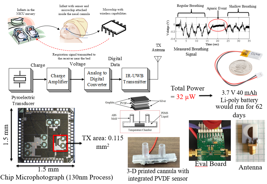
Wireless Respiration Monitoring Sensor for Sleep Apnea
Wireless devices for monitoring respiration activities can play a major role in advancing modern home-based healthcare. Chronic respiratory diseases, such as sleep apnea, are even more critical for premature neonatal infants. PACCAR-affiliated faculty, Prof. Mahbub and her research group, The Integrated Biomedical Circuits and Systems Laboratory (iBioCASL), are designing a low-power wireless respiration monitoring sensor which can reduce the cost and inconvenience of the conventional respiration monitoring systems significantly. At the front-end of the system, a ferroelectric polymer (PVDF) based pyroelectric transducer is utilized to monitor the temperature difference between the room air and the nasal air-flow. The charge generated by the transducer due to this temperature change is then converted to a proportional voltage signal using a low-power low-noise folded-cascode operational transconductance (OTA) based charge amplifier. A pseudo-resistor based diode-connected MOSFET in the feedback configuration is implemented to achieve sub-Hz corner frequency that is required for low-frequency respiration signal monitoring. An apnea detection algorithm has been also been developed in this process and the figure below captures the highlights of this activity.
References:
I. Mahbub, S. Shamsir, S. K. Islam, S. A. Pullano and A. S. Fiorillo, “A low noise front-end amplifier for pyroelectric transducer based respiration monitoring system,” 2017 IEEE International Midwest Symposium on Circuits and Systems (MWSCAS), Boston, MA, 2017.
I. Mahbub, S. K. Islam, S. Shamsir, S. A. Pullano, A. S. Fiorillo, M. S. Gaylord and V. Lorch, "A low power wearable respiration monitoring sensor using pyroelectric transducer," 2017 United States National Committee of URSI National Radio Science Meeting (USNC-URSI NRSM), Boulder, CO, 2017, pp. 1-2.
I. Mahbub M. S. Hasan, S. A. Pullano, F. Quaiyum, C. P. Stephens, S. K. Islam, A. S. Fiorillo, M. S. Gaylord, V. Lorch and N. Beitel, "A low power wireless apnea detection system based on pyroelectric sensor," 2015 IEEE Topical Conference on Biomedical Wireless Technologies, Networks, and Sensing Systems (BioWireleSS), San Diego, CA, 2015, pp. 1-3.
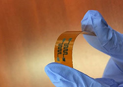
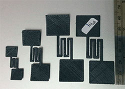
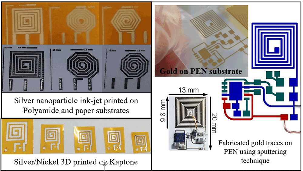
Implementation of Antennas on Flexible Substrates
Prof. Mahbub and her research group, The Integrated Biomedical Circuits and Systems Laboratory (iBioCASL), has designed a wireless power transfer system on various flexible substrates and metal traces, for example using gold on the Polyethylene Naphthalate (PEN), silver ink-jet printed on polyamide and paper and silver/nickel 3D printed on Kapton substrates, as shown in the figure below. Any antenna or device implanted inside the human body is required to have a specific absorption rate (SAR), which is a measure of the rate at which energy is absorbed by the tissue when exposed to a radio frequency (RF) electromagnetic field, to be below 1.67 W/Kg. To verify this, the flexible antennas were designed, and the SAR was simulated using six-layer 10 mm human head tissue model with Ansys HFSS software. Further developments in this area are forthcoming.
Reference:
M. Sinclair, D. K. Biswas, T. Le, J. Hyde, I. Mahbub, L. Change and Y. Hao. “Design of a flexible receiver module for implantable wireless power transfer (WPT) applications” 2019 USNC-URSI National Radio Science Meeting, Boulder, CO, 2019 (under review)
Faculty Affiliated with Focus Area 2
Anupama Kaul
Yuankun Lin
Ifana Mahbub
Marcus Young
Please note, some of the faculty listed above also have overlap activities under Focus Area 1.